Thursday, February 28, 2013

Foreign Spore Germination: Fungi Tumblr

I have shared this tumblr link before but I am sharing it again.

http://mycology.tumblr.com/

Today, because I saw this:
Spider being eaten by fungi

And thought, "Look who weaves the best silken threads now?"

I love that tumblr you should definitely go follow it.

Friday, February 22, 2013

Fungal Word Friday: Columella

The columella is a sterile dome shaped structure found at the tip of a sporangiophore, or within a sporangium.
Rhizopus oryzae with collapsed columella

 Photo Cred: The University of Adelaide Mycology Online: Rhizopus oryzae page.  http://www.mycology.adelaide.edu.au/Fungal_Descriptions/Zygomycetes/Rhizopus/R_oryzae.html

Friday, February 8, 2013

Fungal Word Friday: Adiaconidia

An adiaconidia is a large, globose conidium with a thick wall.
 
Adiaconidia of Emmonsia in scleral nodule biopsy.

Photo Cred: Marcia O. Mendes, Mario A.P. Moraes, Ernesto I.M. Renoiner, Marta H.P. Dantas, Tatiania M. Lanzieri, Carlos F. Fonseca, Expedito J.A. Luna, and Douglas L. Hatch Volume 15, Number 4 issue of Emerging Infectious Diseases. Via Center for Disease Control

Friday, February 1, 2013

Fungal Word Friday: Stolon

The stolon is a running hypha that rhizoids and sporangiospores rise from.
Geomyces destructans pseudo-colored photomicrograph w/ stolon in yellow

It is basically the portion of the hyphae running laterally between the business parts of a fungus.


Photo Cred: Vishnu Chaturvedi, Deborah J. Springer, Melissa J. Behr, Rama Ramani, Xiaojiang Li, Marcia K. Peck, Ping Ren, Dianna J. Bopp, Britta Wood, William A. Samsonoff, Calvin M. Butchkoski, Alan C. Hicks, Ward B. Stone, Robert J. Rudd, Sudha Chaturvedi [CC-BY-2.5 (http://creativecommons.org/licenses/by/2.5)], via Wikimedia Commons

Saturday, January 19, 2013

Fungal Word Friday: Disjunctor cell

First, I apologize that this is on Saturday and not Friday as it should be, I dropped the ball, much as I have been doing of recent weeks on my more in depth pieces. I will find a way to get back on task.

A Disjunctor cell is a cell that breaks open, or undergoes lysis of some form in order to release a conidium.

Coccidioides immitis showing a disjunctor cell on the right.


Photo cred: http://www.studydroid.com/imageCards/03/ef/card-3620318-front.jpg

Friday, January 11, 2013

Fungal Word Friday: Cystidium

Cystidium is a large cell generally seen between clusters of basidia of a basidiomycete. They have a variety of morphological characteristics that can be used to help with species identification. 

Cystidium stained with Congo red


There are several classifications of cystida based on where they grow on the mushroom (on the edge of the lamella, on the face of the lamella, on the margin or surface of the cap, or along the stipe.)
 In addition they can be classified by structure; do they contain a refreactive yellow body (Chrysocystidia), or do they have a granular or oily appearance (Gleocystidia.)

Photo Cred: Peter G Werner (Own image) [CC-BY-SA-3.0 (http://creativecommons.org/licenses/by-sa/3.0)], via Wikimedia Commons

Friday, January 4, 2013

Fungal Word Friday: Hydrophobin

Fungi live all over the place... duh. The thing is, those fungi need to be able to withstand a wide range of environments and pressures in order to do so. On of their tools for survival is a group of proteins called hydrophobins.

Hydrophobin Water Resistant Monolayer assemble!

Hydrophobins are less then 100 amino acids long and rich in cysteine. What makes them special is that only filamentous fungi produce them and they have the ability to self assemble into a monolayer along  hydrophobic:hydrophilic barriers, such as water and air.
Basically hydrophobins give filamentous fungi a barrier to help moderate its interaction with the environment, allowing them to grow in all sorts of habitats.


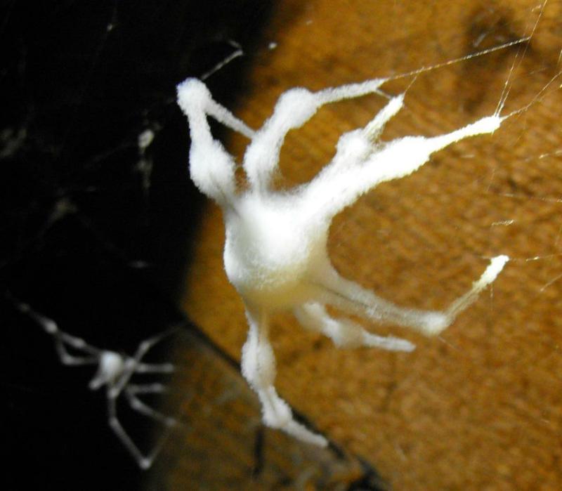
Photo cred:  Lijealso [Public domain or Public domain], via Wikimedia Commons